PASCAR and Boston Scientific joint investment in Sub Sahara Africa - SunEcho Allied Program
Progress on Echocardiography training at Tygerberg Hospital Cardiology Division - Edward Rieth
PROGRESS TO DATE:
As we are all aware that echocardiography is an important non-invasive investigation which provides important information about the structure and function of the heart.
My first encounter with echocardiography started on the 4th April 2016. I was introduced to my tutors and a workplan was laid out. The first two weeks I was bound to the Cath lab, Pacing lab and the Echo section. During this phase I observed the routine echocardiographic examinations that are one of the most common cardiac investigation tools.
Into the third week starting from the 18th April 2016 to the 22nd April 2016 I attended the SunEcho course hosted within the Division of Cardiology in the Faculty of Medicine and Health Sciences of the Stellenbosch University. The course covered a comprehensive syllabus in echocardiography starting from the basic principles to understand an echocardiogram. The course progresses over the five days of which the first day was merely a workshop of 3D-Imaging. The course breakdown was as such:
1st Day: 3D-Workshop
2nd Day: Principles and fundamentals of echocardiography
3rd Day: Evaluating cardiac function and haemodynamics
5th Day: Continuation of Valvular diseases.
It was a comprehensive course that provides a good foundation for the development of skills and encouraged oneself to remain abreast of new development and techniques.
From the fourth week onwards I attended every Tuesday afternoon a lecture on different cardiac topics meant for the cardiac students, physicians, registrars and consultants. On Wednesday afternoons some interesting case studies are presented.
It is now clear that the basic theoretical information has been discussed and now the hands-on assessment becomes the primary objective to detail the transthoracic echocardiograms.
TECHNICAL ASSESSMENT
The standard Two-Dimensional views are obtained from four acoustic windows or transducer locations ( parasternal, apical, subcostal and suprasternal) and three imaging planes ( long axis, short axis and four chambers).
The 2-D examination provides important and detailed information regarding the anatomical structures and the function of the heart.
The motion mode (M-Mode) provides information about the timing of cardiac events.
The Doppler examination provides a graphic display of blood flow velocities plotted over time.
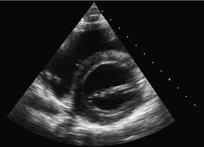
PARASTERNAL SHORT AXIS AT THE LEVEL OF THE AORTIC VALVE
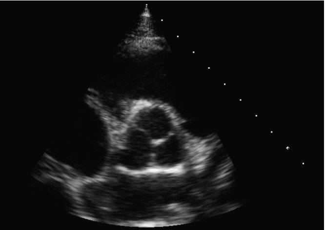
PARASTERNAL SHORT AXIS AT THE LEVEL OF THE MITRAL VALVE
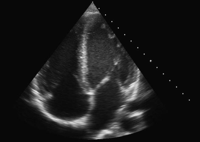
PARASTERNAL LONG AXIS STANDARD VIEW
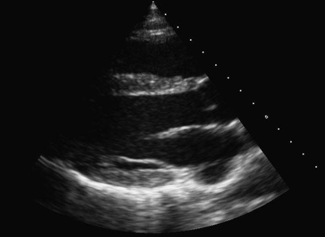
APICAL FOUR CHAMBER VIEW